Return to Slide List
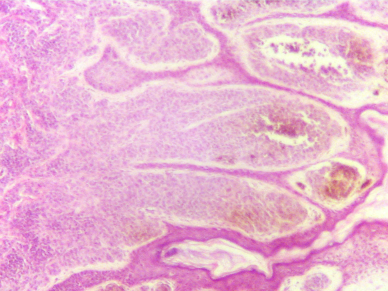
Benign Neoplasm - Intradermal Nevus (Mole)
Note that the stratified squamous epithelium of the epidermis has undergone
proliferation. The epidermis is very thick and contains excessive accumulations
of brown melanin. Compare the thickness of the epidermis in the mole with that
of the normal epidermis at the edges of the section.
Intradermal
nevus (40X1.6)
Normal epidermis (100X2.0)


Dermis (red at bottom), epidermis (blue area and
dark Dermis (red at left),
normal epidermis (dark layer)
layer), air (clear area at right), normal
epidermis (dark
layer in lower region) borders on abnormal thickened
epidermis of nevus (upper blue region with dark surface
layer)
Intradermal nevus (40X1.6)
Intradermal nevus (100X2.0)


Very thickened and rough epidermis (blue with
dark Very
thickened epidermis (blue with dark surface layer) with
surface layer) and brown melanin masses, dermis
(red) brown melanin masses
at extreme bottom
This neoplasm is considered benign if it does not spread but if it invades
into the dermis it would be considered malignant and called a melanoma.
Return to Slide List
Copyright 2020: Augustine G. DiGiovanna, Ph.D., Salisbury University, Maryland
The materials on this site are licensed under CC BY-NC-SA
4.0 .

https://www.biologyofhumanaging.com/Figures/CC-BY-NS-SA%20image.jpg
Attribution-NonCommercial-ShareAlike
This license requires that reusers give
credit to the creator. It allows reusers to distribute, remix, adapt, and build
upon the material in any medium or format, for noncommercial purposes only. If
others modify or adapt the material, they must license the modified material
under identical terms.
Previous print editions of the text Human Aging: Biological Perspectives are ©
Copyright 2000, 1994 by The McGraw-Hill Companies, Inc. and 2020 by Augustine
DiGiovanna.
View License Deed |
View Legal Code


