Return to Slide List

Atherosclerosis of Coronary Artery
 

First observe a slide of an early atherosclerotic lesion. The pink, noncellular material in the intima of the coronary artery is the atherosclerotic plaque. This lesion has already reduced the lumen by about 20%. Now observe one of the later atherosclerotic plaques. Note the degree of occlusion by the lesion. It may take years for plaque to reach this size.   

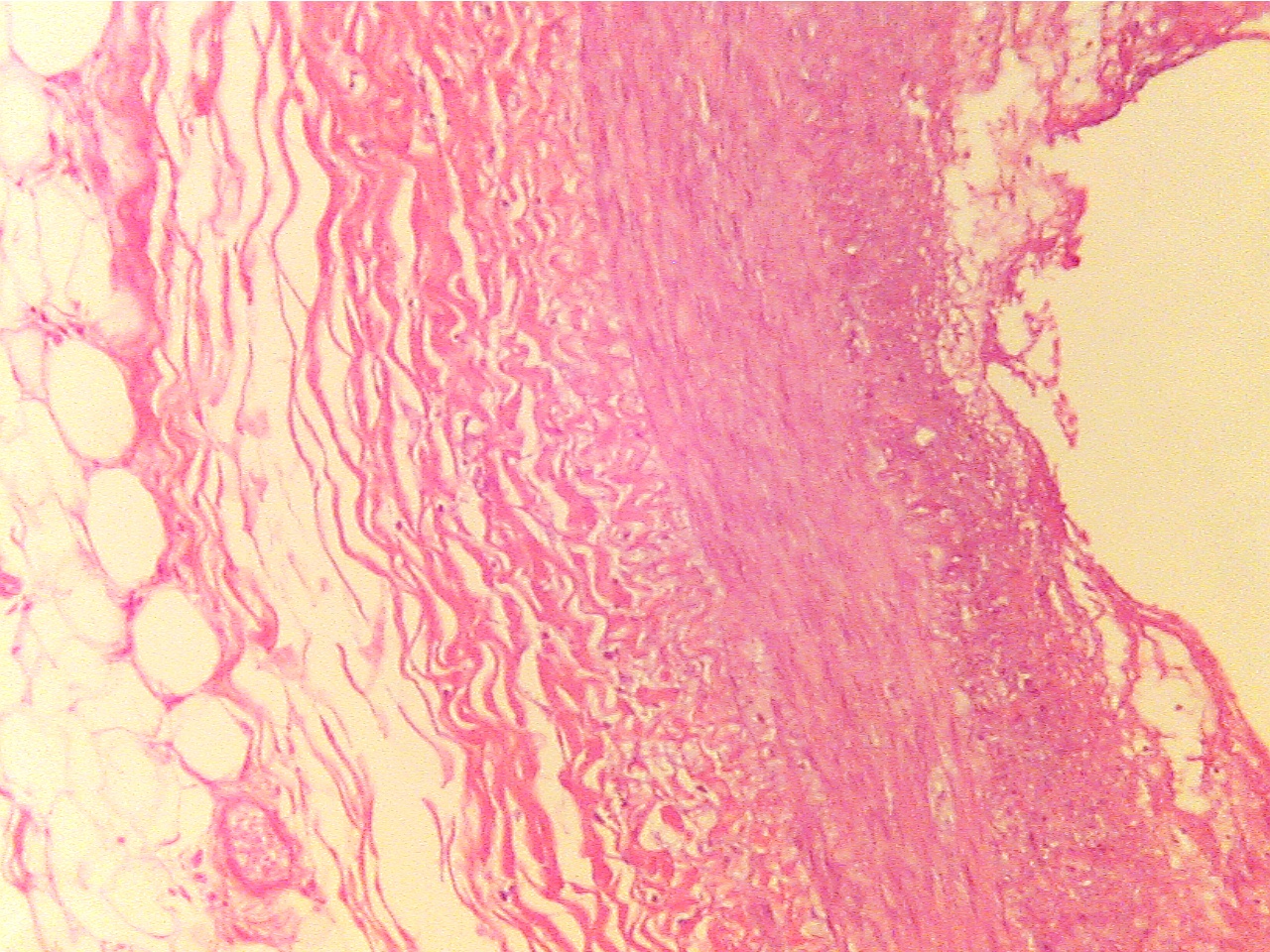
                                          Early lesion (40X1.0 - a1) 
                                       
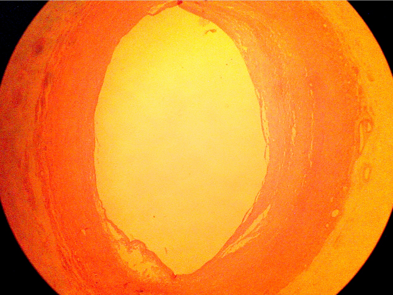
                                        Normal artery wall at left, lumen in center,
                                        lesion (thickened wall) at right. 

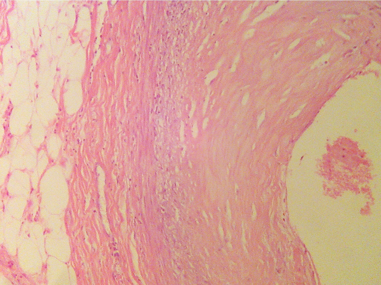
Normal region (100X2.0 - a1)                                                               Early lesion (100X2.0 - a2)
 
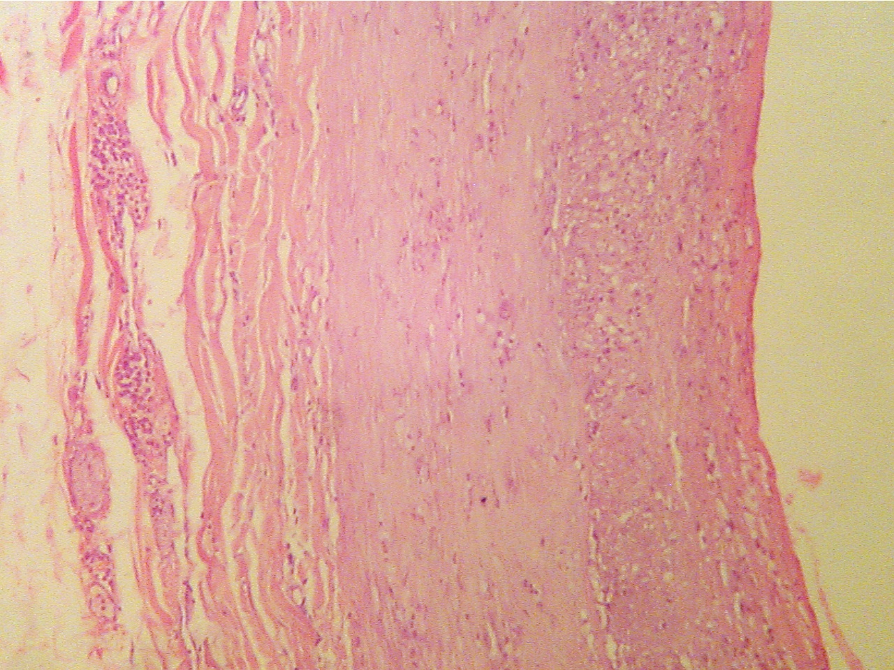
Layers are areolar tissue (left), externa (thick pink                     Lumen at upper left, endothelium (thin layer),
collagen fibers),  smooth muscle (thick middle layer),              intima, fibrous plaque within intima, intima (darker granular
tunica intima (thick granular layer),  endothelium                      layer), smooth muscle (thick pink), externa (lower right)
(thin red layer), lumen at right    

                                          Advanced lesion (40X1.0 - b1)
                                       
                                             Lumen at left of center, lesion at right 

Normal region (100X2.0 - b1)                                                               Advanced lesion (100X2.0 - b2)
 
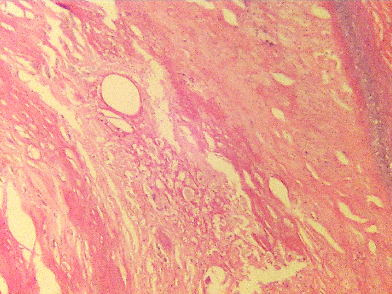
Externa at left, lumen at right                                                     Lumen at far lower left corner, lesion of mixed 
                                                                                                       substances and cells fills center region

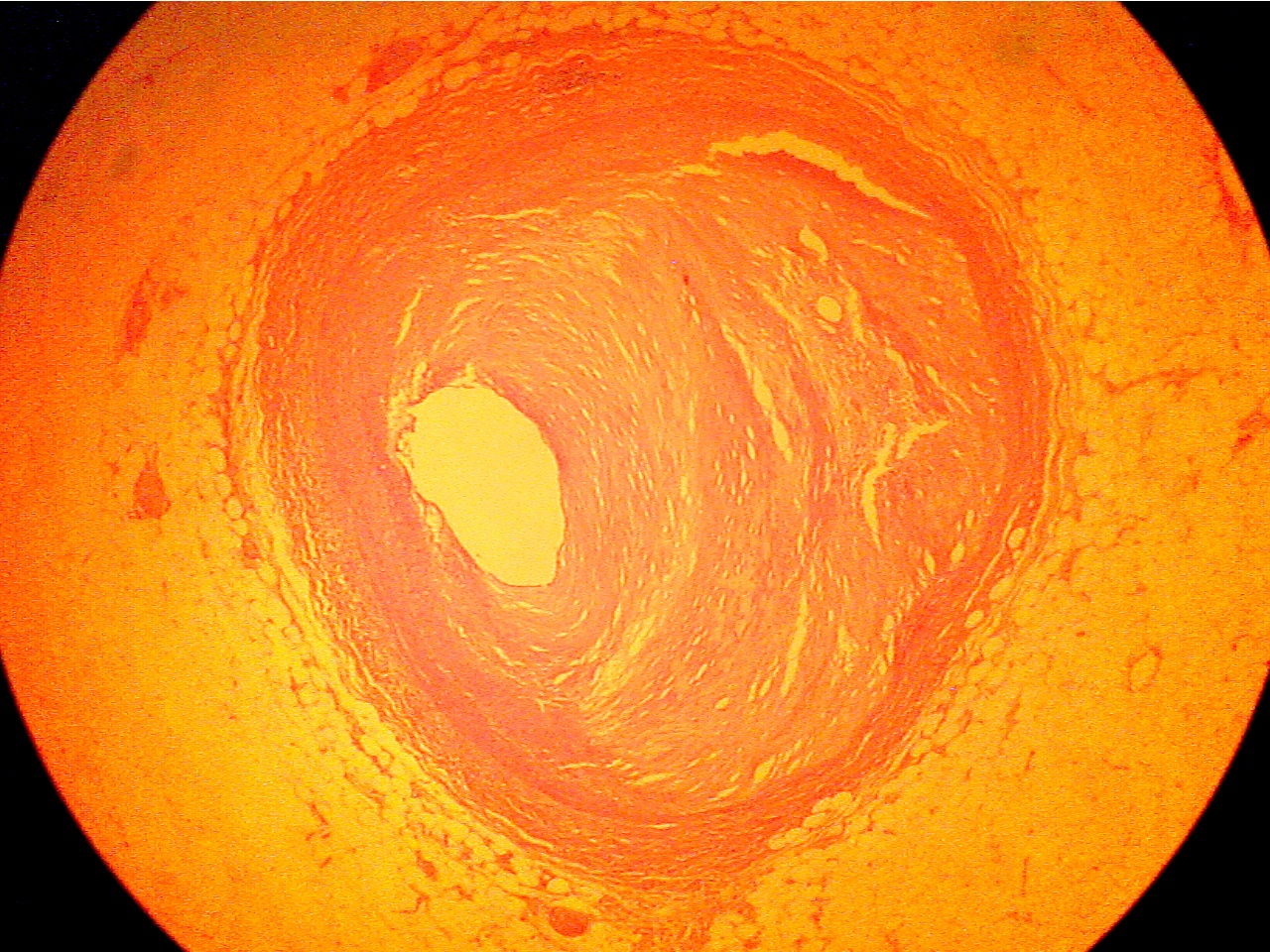
                                            Occlusive lesion (40X1.0 - c1)
                                       
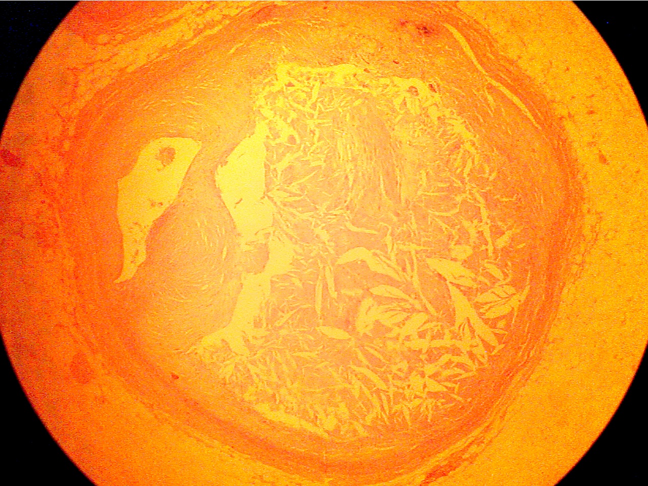
                                          Lumen at far left of center, crystalline lesion fills center

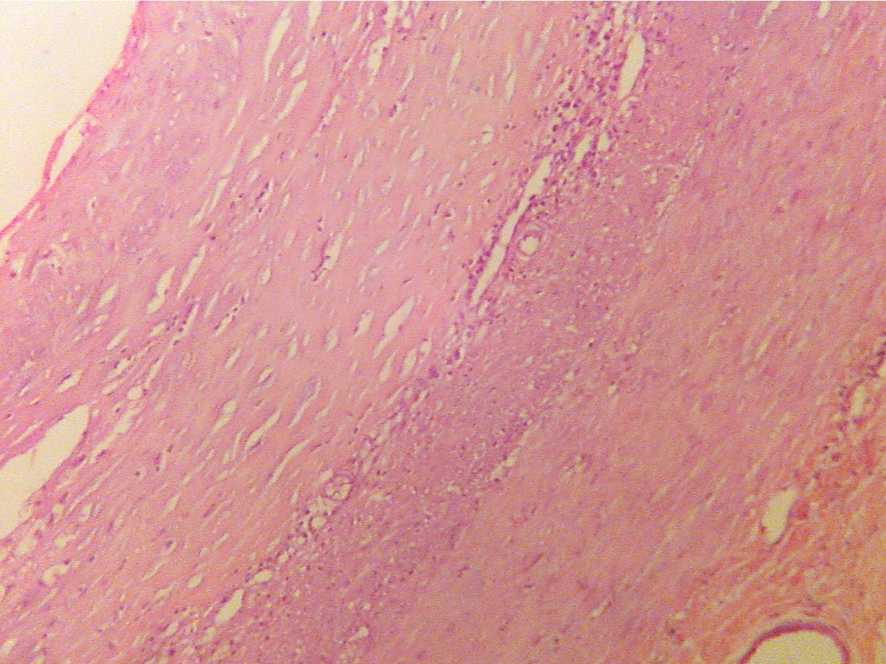
Fibrous plaque (100X2.0 - c1)                                                            Plaque replaces intima and smooth muscle (100X2.0 - c2)
    
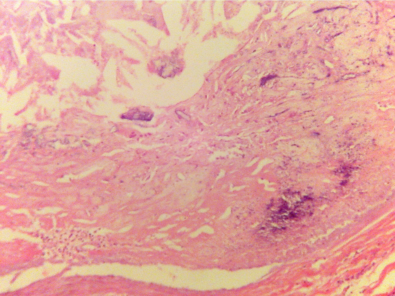
Layers are areolar tissue (left), externa (red collagen              Amorphous acellular material (upper left) and cellular
fibers), smooth muscle (thin red middle layer), tunica           and fibrous plaque (center) replace intima and smooth muscle,
intima (blue granular layer), fibrous plaque (thick, pink),       externa (collagen fibers) remains at lower right
endothelium (thin layer along lumen), lumen (at right)                      

Center of plaque (100X2.0 - c3)                                                          Endothelium (400X2.0 - c4)
  
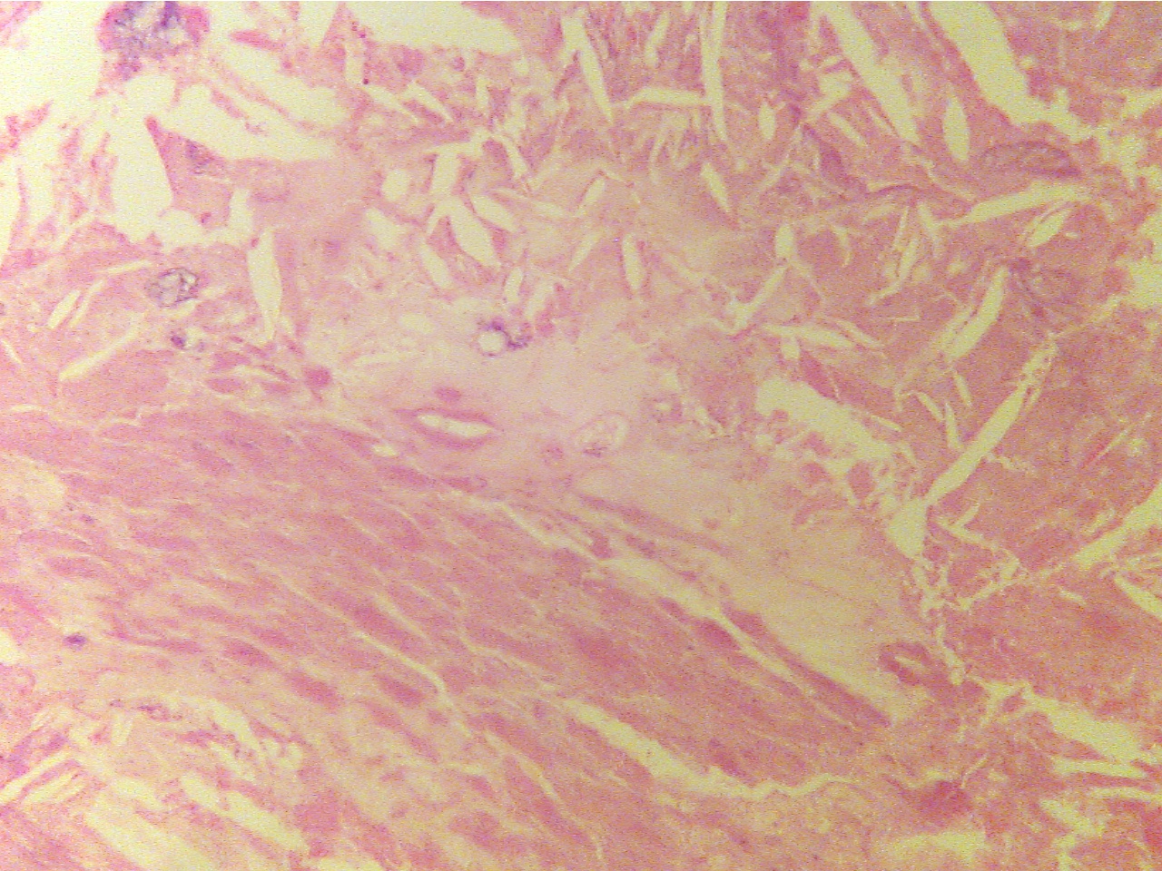
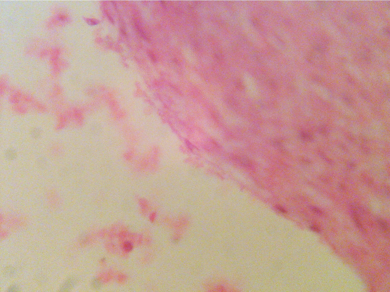
Center of amorphous acellular region                                      Normal endothelium (lower right bordering lumen), plaque 
of plaque, much of which appears crystalline                         (right, underlies endothelium), lumen (left), damaged
                                                                                                        endothelium with adhering RBCs (middle and top) 
Return to Slide List


Copyright 2020: Augustine G. DiGiovanna, Ph.D., Salisbury University, Maryland

The materials on this site are licensed under CC BY-NC-SA 4.0 .


 https://www.biologyofhumanaging.com/Figures/CC-BY-NS-SA%20image.jpg
Attribution-NonCommercial-ShareAlike

This license requires that reusers give credit to the creator. It allows reusers to distribute, remix, adapt, and build upon the material in any medium or format, for noncommercial purposes only. If others modify or adapt the material, they must license the modified material under identical terms.
Previous print editions of the text Human Aging: Biological Perspectives are © Copyright 2000, 1994 by The McGraw-Hill Companies, Inc. and 2020 by Augustine DiGiovanna.

View License Deed | View Legal Code